What is Uterine Septoplasty/Metroplasty
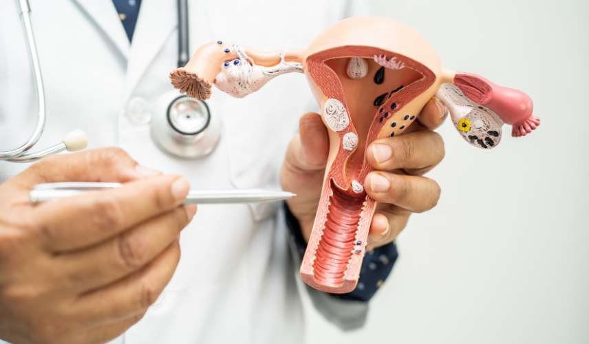
A uterine septum is a common anomoly of the uterus that is seen in 1 – 15 per 1,000 women. In this condition, the cavity of the uterus is separated by a long piece of tissue, while the outside of the uterus has a normal shape. In a uterine septum, a wall or wedge of tissue divides the uterus; it is composed of muscle fibers and some connective tissue. It may be complete, dividing the uterus into completely separate chambers, or partial. The size and shape of the septum can vary by width, length, and blood supply.
How is a uterine septum diagnosed?
The gold standard method requires direct visualization of the exterior and interior of the uterus using laparoscopy and hysteroscopy. These are relatively invasive and currently, imaging techniques are the preferred modality to diagnose uterine septum. Use of X-ray (hysterosalpingography) is often the initial test to diagnose an anomaly. However, it is not possible to differentiate between a septate uterus and a bicornuate uterus (double uterus) with X-ray. Use of ultrasound (hysterosonogram or sonohysterogram) is a good technique for diagnosing a uterine septum. Saline is injected in the uterine cavity with concurrent vaginal ultrasound scanning. This enables one to visualize the inner as well as the outer contour of the uterus. 3-dimensional (3-D) ultrasound combined with saline infusion has 100% accuracy when compared with surgery. Magnetic Resonance Imaging (MRI) can also be used for diagnosis of uterine septum.
